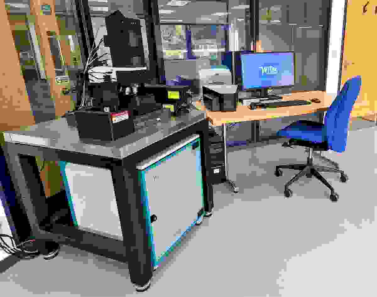
Since we welcomed WITec into the Oxford Instruments family, we had been looking forward to developing our Raman spectroscopy lab. So, we were very excited about the recent installation of a WITec alpha300R Raman microscope in our High Wycombe laboratories and the new complementary capabilities it provides us.
A quick introduction to me
I have recently joined Oxford Instruments as a Product Scientist focusing on the exciting combination of Raman and Electron Microscopy based analysis. I have come fresh from completing a postdoctoral position and my PhD, both of which focused on the use of Raman spectroscopy for materials analysis. I have worked across a wide range of applications, including biological samples to energy storage materials, and am now hoping I can apply myself and my experience successfully to this new role.
Raman at a glance
I won’t be going into too much detail here but do look out for future posts where I will be discussing the technique in much greater detail. Raman microscopy offers a different but complementary perspective to Oxford Instruments’ NanoAnalysis capabilities. It can deliver high-quality Raman imaging in both 2- and 3-dimensions using a confocal optical microscope. Exciting our molecule with laser light, we can obtain insightful chemical and structural data about the molecules that make up our material. We can also distinguish polymorphs via the detection of phonons from crystalline structures. This enables us to generate highly resolved structural maps relating to changes in the composition of our material. This does require practically perfect alignment of the internal components of our microscope. I can assure you that this can be a painstaking process and patience is required!

A photograph of our new WITec alpha300R Raman microscope here at High Wycombe
Our system
We have some incredibly smart features with our alpha300R system that I personally cannot wait to play around with and really test the limits of the instrument – Again, do look out for future posts where I will dive a little deeper!

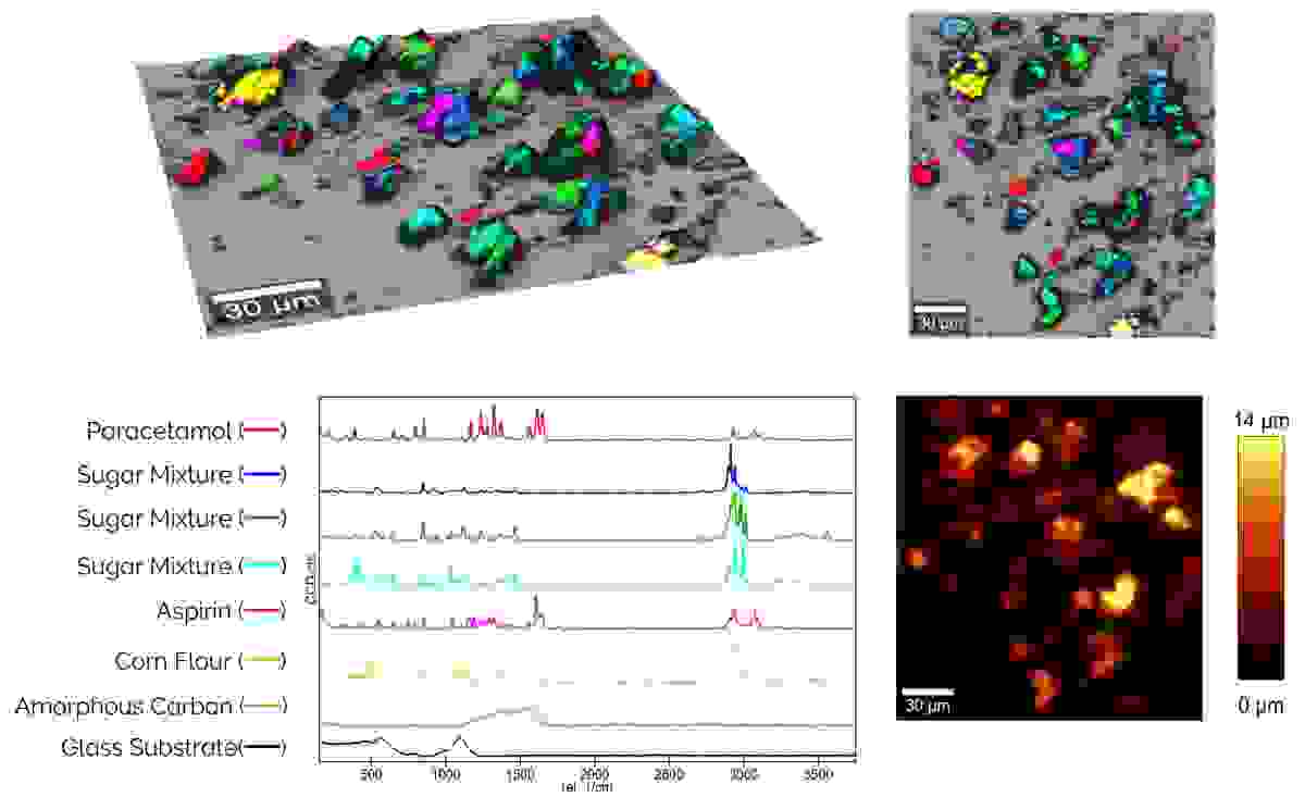
The first Raman study conducted here at High Wycombe, identifying different components of a powder mixture comprising of a common painkiller and sugar.
This includes the WITec TrueSurface and ParticleScout packages. TrueSurface gives us the ability to create topological data alongside our 2D Raman image. Using a cleverly designed optical profilometer, we can keep our sample in focus during large area mapping regardless of the topographical variation. This, combined with the excellent spatial resolution of the confocal microscope means we can create incredibly detailed Raman maps. ParticleScout allows us to conduct automated, high-quality particle analysis with the ability to distinguish the individual components of our powder. We can also categorise our particles by numerous parameters, including size and shape, producing in-depth reports that summarise our results.
This WITec alpha300R Raman microscope is a fantastic addition to our demonstration facility here at High Wycombe. Now, the real fun can begin and I hope that this blog has enticed you to learn more. I will be back soon with more posts that will really focus in on the core principles and capabilities of the system, and I hope you will join me on this journey.


