Raman microscopy is rapidly evolving from specialist technique into standard kit for research laboratories. The number of fields that use it, and the variety of its application within those disciplines, is steadily increasing. Advances in user-friendliness and experimental versatility are the primary factors driving this expansion.
Following its discovery and experimental demonstration by C.V. Raman in 1928, Raman spectroscopy first became practical with the advent of several revolutionary photonics devices. Lasers – developed in the 1960s – provided monochromatic light in a range of wavelengths for sample excitation, and charge-coupled devices – first commercially available in the 1980s – offered sufficient sensitivity to acquire large numbers of Raman spectra quickly. These technologies initiated the first substantial wave of adoption, primarily among physicists, chemists, and materials scientists.
Fig.1: A replica of the original gas tube and ruby rod laser, invented by Theodore Maiman (left) and a picture of a charge-coupled device (right).
The 1990s brought more powerful personal computers and precise piezo-driven sample scanning stages that enabled instruments to acquire a complete Raman spectrum at each measurement point and compile them to generate an image. WITec GmbH introduced Raman imaging to the marketplace in 1999 with the first generation alpha300 microscope. Once scientists saw high-resolution 3D visualisations of sample component distributions, demand for the technique quickly spread.
Fig. 2: The Apple iMac, released in 1998 (left) and the first edition of the WITec alpha300 Raman microscope (right).
Today Raman imaging is found far beyond its initial user base, in archaeology, life science, nutrition, forensics, pharmaceutical development, geoscience and many other fields that require detailed, label-free, nondestructive analysis.
Advanced control software and motorised optical elements have made even complex investigations easily manageable. Automation removes the need for manual fine adjustment and calibration while aiding reproducibility and eliminating potential sources of error. This benefits all researchers, from university groups and multi-user facilities to industrial quality control teams. WITec’s alpha300 apyron represents the current state of the art in fully automated Raman microscopes.
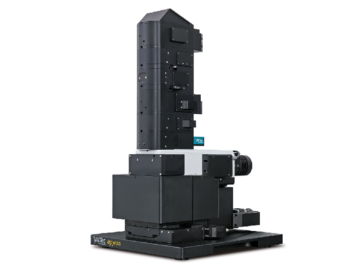
Fig. 3: Automated Raman imaging system, Oxford Instruments WITec alpha300 apyron.
The future of Raman microscopy will see the flourishing of new forms of correlative analysis and expanded options for sample handling and access. Some recent advances include portable coordinate systems for relocating areas of interest across multiple platforms and configurations designed around distinct sample forms.
WITec’s modular alpha300 systems can already incorporate fluorescence imaging, SHG/THG, SNOM, AFM and optical profilometry, while RISE (Raman Imaging Scanning Electron) Microscopes integrate Raman and SEM within a common vacuum chamber. ParticleScout brings Raman analysis to microparticle and microplastic samples and cryoRaman can examine materials near absolute zero where quantum effects are more apparent. The alpha300 Semiconductor Edition was made to accommodate up to 300 mm wafers and the alphaCART mobile Raman system removes the size limit on samples altogether by allowing researchers to take their laboratory on-site.
Fig. 4: RISE Microscopes: Fully-integrated Raman Imaging and Scanning Electron Microscopy
The evolution of Raman spectroscopy – from Nobel Prize-winning experiment, to expert’s choice for chemical characterisation, toward becoming a standard technique, and moving beyond single method analysis and past conventional sample formats – has been a series of steps toward fulfilling its initial promise. Now that everyone has access, the research community is devising novel ways to apply it to emerging challenges. Raman microscopy, in all its forms, looks set to become ubiquitous in laboratories around the world and across the full range of scientific inquiry.